Geometric deep learning and graphical models for accurate and traceable medical image analysis
Deep Learning, as a weak form of Artificial Intelligence, has already led to major advances in various areas of medical image analysis, such as the fully automated segmentation of anatomical structures in CT and MRI image volumes. It has the potential to become the most important component for modern medical image understanding. At the same time, there are still a number of major challenges in deep learning based image processing, such as the high sensitivity to intensity changes (e.g. due to different scanner types) or the increasingly complex network structures whose decision making is difficult to understand for humans.
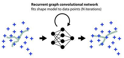
In this research project it will be investigated to what extent anatomical prior knowledge (e.g. about automatically extracted landmarks) and graphical models can be integrated into deep neural networks to improve their performance and explainability. In particular, methods from the field of Geometric Deep Learning are used, which can explicitly include geometries and learned feature vectors of investigated structures with the help of graph-based neural networks.
In order to clarify the general benefit of the developed methods, they are applied and evaluated in various classical areas of medical image processing. These include anatomical landmark detection, semantic segmentation of organ surfaces and the registration of severely deformed 4D volumes (e.g. thorax CT during inhalation and exhalation).
Selected Publications
- Hansen L., Heinrich M.P.
Sparse Structured Prediction for Semantic Edge Detection in Medical Images
International Conference on Medical Imaging with Deep Learning (MIDL, 2019)

- Research
- AI und Deep Learning in Medicine
- Medical Image Processing and VR-Simulation
- Integration and Utilisation of Medical Data
- Sensor Data Analysis for Assistive Health Technologies
- Medical Image Computing and Artificial Intelligence
- Medical Data Science Lab
- Medical Deep Learning Lab
- Junior Research Group Diagnostics and Research of Movement Disorders
- Former Medical Data Engineering Lab