JOINT: Joint Disorders in Paediatric Orthopaedics with Interactive 3D Ultrasound Navigation – Human + Technology
Hip dysplasia is the most common congenital skeletal anomaly worldwide, with an incidence of 2–4%. If left untreated, it can lead to osteoarthritis and necessitate joint replacement at an early age. In newborns, however, hip dysplasia can be fully corrected through non-surgical measures if detected early. In Germany, screening is therefore part of the statutory U-examinations, during which both hip joints are assessed by ultrasound to determine the degree of femoral head coverage by the acetabulum.
This ultrasound examination is demanding even for experienced clinicians and is therefore highly operator-dependent. Traditionally, a specific sectional view is sought, in which various hip angles can be measured according to the Graf method. The aim of the JOINT project is to simplify and enhance this examination through the use of artificial intelligence. By recording sweeps rather than individual sectional images, three-dimensional reconstruction of the ultrasound scans becomes possible. This enables precise 3D reconstruction of hip anatomy using affordable and radiation-free ultrasound diagnostics.
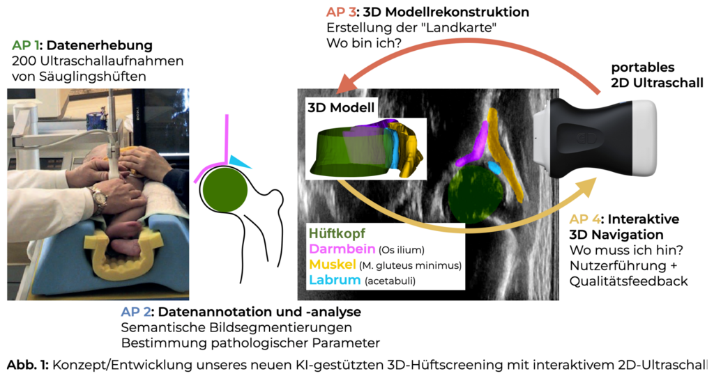
From these reconstructions, artificial intelligence can not only identify the Graf plane and calculate the required angles, but also exploit the three-dimensional representation to discover and apply novel, potentially more accurate markers and metrics for diagnosing dysplasia. On this basis, the AI can be trained to recognise pathological findings. The project therefore aims to develop methods that support less experienced clinicians in diagnosis and improve neonatal screening.
In addition to the University of Lübeck, the project involves EchoScout GmbH and the University Medical Centre Schleswig-Holstein (UKSH), Kiel.
Funded by the BMFTR (2024–2026) with 500,372€ (79,800€ allocated to IMI).
Selected Publications:
Heinrich, Mattias P., et al. "Chasing clouds: differentiable volumetric rasterisation of point clouds as a highly efficient and accurate loss for large-scale deformable 3D registration." Proceedings of the IEEE/CVF international conference on computer vision. 2023.
Heyer, Wiebke, et al. "Autocalibration for 3D Ultrasound Reconstruction in Infant Hip Dysplasia Screening." BVM Workshop. Wiesbaden: Springer Fachmedien Wiesbaden, 2025.
Hansen, Lasse, and Mattias P. Heinrich. "Deep learning based geometric registration for medical images: how accurate can we get without visual features?." International conference on information processing in medical imaging. Cham: Springer International Publishing, 2021.
Project Team:
- Research
- AI und Deep Learning in Medicine
- Medical Image Processing and VR-Simulation
- Integration and Utilisation of Medical Data
- Sensor Data Analysis for Assistive Health Technologies
- Medical Image Computing and Artificial Intelligence
- Medical Data Science Lab
- Medical Deep Learning Lab
- Junior Research Group Diagnostics and Research of Movement Disorders
- Former Medical Data Engineering Lab
Contact person
Mattias Heinrich

Professor
Gebäude MFC2, 4.OG
mattias.heinrich(at)uni-luebeck.de
+49 451 3101 5602
Wiebke Heyer

Research Assistant
Gebäude MFC2, 4.OG
w.heyer(at)uni-luebeck.de
+49 451 3101 5609