Finite Element Modeling of Respiratory Lung Motion
Problem
Respiratory motion is a main source of error in radiation therapy of thoracic tumors. In current clinical practice treatment planning is usually based on 3D CT imaging. Organ and tumor motion is accounted for by increasing safety margins which in turn increases the volume of healthy tissue to be irradiated. Development and optimization of methods to adequately account for breathing motion require detailed knowledge about respiratory dynamics. Consequently, computer aided modeling and model based simulation gain in importance. Within this project we aim at modeling respiratory (lung) motion taking the physiology of breathing as modeling starting point. As an approved method in biophysical model-ing we therefore apply Finite Element Methods (FEM). Since FEM enable to directly model certain (bio-) physical aspects by means of e.g. boundary conditions it provides for a high-precision analysis and simulation.
Methods
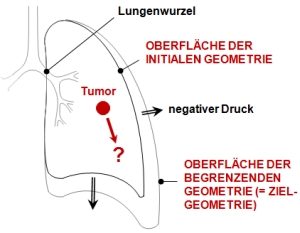
Starting point of modeling is the process of lung ventilation. During breathing the thoracic cavity is expanded by contraction of the diaphragm and outer intercostal muscles. This causes changes in the intrapleural pressure which acts as a force upon the lung surface: Lung expands, and (since the pleu-ral cavity is filled with a fluid) during this process the visceral pleura is sliding frictionlessly down the internal surface of the thoracic cavity.
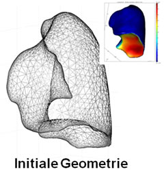
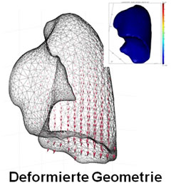
This process is modeled by means of Finite Element Methods: A uniform negative pressure is applied to a lung surface representing some initial state of breathing (e.g. state of end-expiration). This causes the lung to expand. We limit expansion by a geometry representing the lung shape at a final state of breathing (e.g. end-inhalation); see fig. 1 for illustration. If there occurs a contact between the two geometries, the contact is modeled to be frictionless. For simplistic purposes lung tissue is assumed to be an isotropic linear elastic and homogeneous medium. As FEM software we use COMSOL Mul-tiphysics (FEMLAB, Sweden).
The modeling approach enables to generate patient specific models. Therefore we use 4D CT image data with high spatial and temporal resolution to create such models. Modeling accuracy is evaluated comparing simulated patient specific motion patterns of inner lung landmarks and corresponding mo-tion patterns observed in the 4D CT data. The influence of biomechanical and geometrical parameters (elasticity modulus, Poisson’s ratio; mesh quality) on the modelling process and modelling accuracy is evaluated.
Selected Publications
- René Werner, Jan Ehrhardt, Rainer Schmidt, Heinz Handels
Patient-Specific Finite Element Modeling of Respiratory Lung Motion using 4D CT Image Data
Medical Physics, 36, 5, 1500-1511, 2009. - René Werner, Jan Ehrhardt, Rainer Schmidt, Heinz Handels:
Modeling Respiratory Lung Motion: A Biophysical Approach using Finite Element Methods
In: Hu X.P., Clough, A.V. (eds.), Physiology, Function, and Structure from Medical Images, SPIE Medical Imaging 2008, San Diego, Vol. 6916, 0N-1-0N-11, 2008.
Project Team
Dipl.-Inf. Dipl.-Phys. René Werner
Dr. Jan Ehrhardt
Prof. Dr. Heinz Handels
- Research
- AI und Deep Learning in Medicine
- Medical Image Processing and VR-Simulation
- Integration and Utilisation of Medical Data
- Sensor Data Analysis for Assistive Health Technologies
- Medical Image Computing and Artificial Intelligence
- Medical Data Science Lab
- Medical Deep Learning Lab
- Junior Research Group Diagnostics and Research of Movement Disorders
- Former Medical Data Engineering Lab