Automatic Analysis and Recognition of Image Structures in 3D/4D OCT Image Data using Machine Learning Methods
Optical Coherence Tomography (OCT) is a non-invasive imaging technique based on light of short coherence length. OCT works on the principle of ultrasound imaging, using light instead of sound waves. Interferometry is used to measure differences in transit time between the backscattered light of the sample beam and the light of the reference beam, which allows three-dimensional volume images with micrometer resolution.
Deep learning methods for improved monitoring of age-related macular degeneration in time-spatial 4D-OCT image sequences
In this project deep learning based learning methods for an improved monitoring of age-related macular degeneration (AMD) are developed. The project is carried out in cooperation with the Clinic for Ophthalmology of the University Medical Center Schleswig-Holstein in Kiel and the Institute for Biomedical Optics of the University of Lübeck.
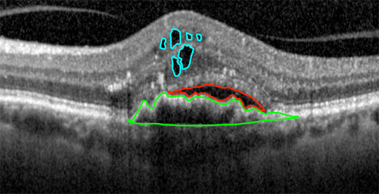
Age-related macular degeneration (AMD) is the most common cause of blindness at the age of 60 and older in the western world. In the wet, exudative form of AMD an edema formation under the retina occurs. Due to an oxygen deficiency in the photoreceptors, messenger substances such as the Vascular Endothelial Growth Factor (VEGF) are formed, which leads to choroidal neovascularization. The newly formed vessels are very fragile, causing unwanted bleeding in the macula. In order to counteract the uncontrolled blood vessel growth, VEGF antibodies can be injected into the vitreous cavity. However, the therapeutic effect only lasts for a few weeks, so that a Reiinjektion is necessary after this period. To improve the diagnosis and therapy of AMD, the project will develop, implement and validate new deep learning methods for computer-aided analysis and detection of biomarkers (Fig. 1) in spatio-temporal 4D-OCT image data. The developed methods are intended to detect and classify the biomarkers in the OCT image data so that a recurrence can be detected early.
Machine Learning Methods for Segmentation and Analysis of the Subcutaneous Fat Layer of Mouse Skin in 3D/4D-OCT Image Data
The aim of this project is the automatic segmentation and quantitative analysis of subcutaneous adipose tissue of mice in 3D-OCT image data using machine learning methods, which are needed for the evaluation of the cryolipolysis procedure. This project is carried out in collaboration with the Cutaneous Biology Research Center at Massachusetts General Hospital in Boston.
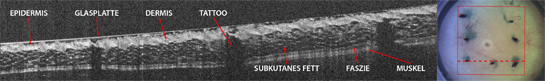
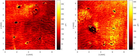
Cryolipolysis is a cosmetic procedure for non-invasive fat reduction co-developed by the cooperation partner Dr. Dieter Manstein. Using a special controlled cooling technique, subcutaneous fat cells can be selectively destroyed. A mouse model is to be tested for the future evaluation of the procedure. Using OCT, 3D images of the mouse skin can be obtained with micrometer resolution (Fig. 1). This high resolution results in a large amount of image data, the manual evaluation of which is practically impossible due to the high expenditure of time. The aim of this project is to develop a computer-aided method to automatically generate segmentations of the subcutaneous fat layer in 3D-OCT image data (Fig. 2). For the implementation, learning-based methods with high accuracy and robustness will be developed.
Selected Publications
- Kepp T., Droigk C., Casper M., Evers M., Salma N., Manstein D., Handels H.,
Segmentation of subcutaneous fat within mouse skin in 3D OCT image data using random forests", Proc. SPIE 10574, Medical Imaging 2018: Image Processing, 1057426 (2 March 2018); doi: 10.1117/12.2290085 - Kepp T., Droigk C., Casper M., Evers M., Salma N., Manstein D., Handels H.
Abstract: Random-Forest-basierte Segmentierung der subkutanen Fettschicht der Mäusehaut in 3D-OCT-Bilddaten, In: Maier A., Deserno T.M., Handels H., Maier-Hein K.H., Palm C., Tolxdorff T. (eds.), Bildverarbeitung für die Medizin 2018, Erlangen, Informatik aktuell, Springer Vieweg, Berlin Heidelberg, 203, 2018
Project Team
M.Sc. Timo Kepp
Prof. Dr. rer. nat. habil. Heinz Handels (Head)
Cooperation Partners
PD Dr. rer. nat. Gereon Hüttmann
Institut für Biomedizinische Optik
Universität zu Lübeck
Dr. med. Claus von der Burchard
Klinik für Ophthalmologie
Universitätsklinikum Schleswig-Holstein, Kiel
Prof. Dr. med. Johann Roider
Klinik für Ophthalmologie
Universitätsklinikum Schleswig-Holstein, Kiel
Dr. Dieter Manstein
Cutaneous Biology Research Center
Massachusetts General Hospital, Boston, Massachusetts
- Research
- AI und Deep Learning in Medicine
- Medical Image Processing and VR-Simulation
- Integration and Utilisation of Medical Data
- Sensor Data Analysis for Assistive Health Technologies
- Medical Image Computing and Artificial Intelligence
- Medical Data Science Lab
- Medical Deep Learning Lab
- Junior Research Group Diagnostics and Research of Movement Disorders
- Former Medical Data Engineering Lab