Artefact reduced Reconstruction of 4D-CT Data Sets from 3D-CT Data Segments
Problem
Modern CT scanners are able to acquire multiple slices at the same time. But the volume scanned within one rotation of the gantry is very often too small to image organ motion. But applications e.g. in radiation oncology may need information about organ motion. It is for example important to image and measure organ motion caused by breathing. For this reason tumour patients have been scanned several times with a CT scanner delivering single 3D-CT data segments. With the use of these segments 4D-CT data sets can be reconstructed. They can be used for further analysis of organ motions due to breathing.
Methods
A modern multi-slice CT scanner has been used to scan a part (= segment) of the patients during free breathing [1]. The position within the breathing cycle was measured with a spirometer so that multiple CT data segments were acquired at different positions within the breathing cycle. By moving the CT couch data segments for the whole thoracic region could be scanned.
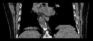
The 3D-CT data segments have to be processed to reconstruct a 4D-CT data set from the 3D-CT data segments. Therefore data segments are needed for each couch position that have been acquired in the same position within the breathing cycle. Using standard techniques, segments are selected that are closest to the lung volume chosen (fig. 1). This method can be called "nearest neighbour interpolation". Segments that are most similar to the volume desired are chosen. By the use of this method artefacts occur that are visible especially at the diaphragm.
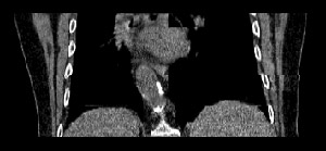
To reduce these artefacts an interpolation method has been developed using non-linear image registration methods. The non-linear registration method is based on the "optical flow". The application of this method on two data segments approximates a vector transformation field describing the shift of the single voxels between neighbouring lung volumes. With this 3D transformation field data segments can be calculated for the lung volume selected. The results (fig. 2) show less artefacts than the images reconstructed with the nearest neighbour strategy (fig. 1).
Selected Publications
- Jan Ehrhardt, René Werner, Thorsten Frenzel, Dennis Säring, Wei Lu, Daniel Low, Heinz Handels: Optical Flow based Method for Improved Reconstruction of 4D CT Data Sets Acquired During Free Breathing.
Medical Physics, 34, 2, 711-721, 2007. - René Werner, Jan Ehrhardt, Thorsten Frenzel, Dennis Säring, Wei Lu, Daniel Low, Heinz Handels: Motion Artifact Reducing Reconstruction of 4D CT Image Data for the Analysis of Respiratory Dynamics.
Methods of Information in Medicine, 46, 254-260, 2007. - Heinz Handels, René Werner, Thorsten Frenzel, Dennis Säring, Wei Lu, Daniel Low, Jan Ehrhardt: Generation of 4D CT Image Data and Analysis of Lung Tumour Mobility During the Breathing Cycle.
Stud Health Technol Inform, 124, 977-982, 2006.
Project Team
Dr. Jan Ehrhardt
Dipl.-Inf. Dipl.-Phys. René Werner
Prof. Dr. Heinz Handels
Cooperation Partners
Prof. Daniel Low and Dr. Wei Lu
Mallinckrodt Institute of Radiology
Washington University
St. Louis, USA
Dr. med. Dr. rer. nat. Thorsten Frenzel
Klinik und Poliklinik für Strahlentherapie und Radioonkologie
Universitätsklinikum Hamburg-Eppendorf

- Research
- AI und Deep Learning in Medicine
- Medical Image Processing and VR-Simulation
- Integration and Utilisation of Medical Data
- Sensor Data Analysis for Assistive Health Technologies
- Medical Image Computing and Artificial Intelligence
- Medical Data Science Lab
- Medical Deep Learning Lab
- Junior Research Group Diagnostics and Research of Movement Disorders
- Former Medical Data Engineering Lab