Decision Forest Variants for Brain Lesion Segmentation
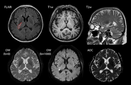
Many diseases, such as ischemic stroke and multiple sclerosis (MS), cause focal alteration of the brain tissue, so called lesions, which are visible in magnetic resonance (MR) imaging sequences (see Fig. 1). Reliable diagnosis and informed treatment decisions require a quantitative assessment of these lesions in time and space, which can only be obtained through further analysis of the imaging data. A brain lesion segmentation that meets the high requirements on the accuracy, reproducibility and robustness placed by clinical and research standards, is in high demand.
In this project, new methods for brain lesion segmentation based on decision forests (DF) were developed and investigated.
Brain lesion segmentation in multi-spectral MR images with decision forests
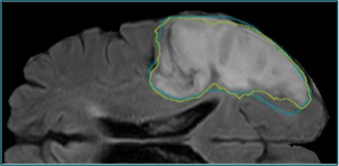
By intelligently combining a chain of MR preprocessing methods, a set of carefully handcrafted image features and the robust DF classifiers, a novel automatic method for the exact and robust segmentation of brain lesions from multi-spectral MR images was developed. The solution is tailored towards a use in clinical practice and research scenarios and as such poses minimal requirements on the quality of the input data. Since it follows the machine learning paradigm, the method can be adapted to various types of brain lesion causing diseases by simple re-training and has been accordingly evaluated on stroke (see Fig. 2), MS and glioma cases. The approach achieved high ranks and won multiple awards on international benchmarks on brain lesion segmentation stoke (ISBIMS2015, ISLES2015, BRATS2015, ISLES2016), proving its outstanding segmentation performance and high adaptability to new diseases.
Local problem forests
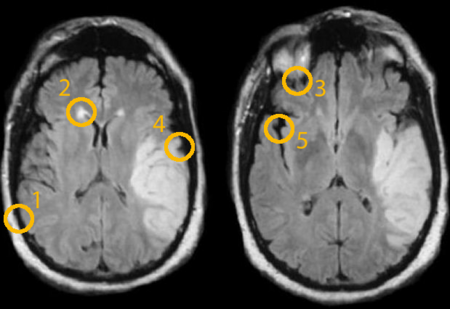
The segmentation of brain lesions from MR images incorporates a number of particular challenging areas (see Fig. 3). To target these spatially local problems, a methodological extension of the DF models was developed. By preceding the training of the forest’s trees by an unsupervised spectral clustering step based on image patches, a topology of local segmentation problems is created. Instead of using bagging, trees are placed in the topology’s areas of high training patch accumulations and trained on the proximal training samples with a normal distributed fuzzy catchment area. Thus, trees specializing on particular subproblems are trained and selectively applied to new cases, significantly increasing the segmentation accuracy for stroke segmentation from mono-spectral MR images.
Semi-supervised forests
In particular for MS, the lesion dissemination in time is as important as in space and MR scans are regularly conducted to monitor disease progression. Based on the manual segmentation of one time point’s scans, the previously developed classification method can be trained and used to segment the subsequent time points automatically. But, following the semi-supervised segmentation paradigm, it is potentially beneficial to incorporate the unlabeled testing samples from the time point to be segmented in the training process driven by the labeled samples from previous time points.
To this end, the DF model was methodologically extended to a allow for training with partially labeled training data. While the standard DF optimize the information gain via the Shanon entropy to determine the best split at each tree node, this novel approach balances the labeled term against a term based on differential entropy representing the unlabeled samples. Thus, each split is set at the equilibrium between label purity and cluster density in feature space.
Applied to longitudinal MS segmentation, the proposed semi-supervised forest lead to a significant improvement in segmentation accuracy when compared to the classical supervised DFs.
Summary
In the scope of this project, a robust and accurate brain lesion segmentation method was developed and shown to outperform the state of the art. Additional methodological and algorithmic improvements designed for specific use-cases furthermore improved the segmentation accuracy. All methods were evaluated on brain MR data of clinical relevance in direct international benchmarks and are currently employed in research settings.
The project was carried out in close cooperation with experts from the cognitive neuroscience group at the Department of Neurology, University Medical Center Schleswig-Holstein, Germany. Oskar Maier is a member of the Graduate School for Computing in Medicine and Live Science, Universität zu Lübeck, Germany.
Code
MedPy – Medical Image Processing in Python
package (PyPI) / source code (GitHub)
sklearnef – Extension module providing un- and semi-supervised decision forests for scikit-learn
source code (GitHub)
DynStatCov - Cython Library for fast dynamic statistical co-variance update
package (PyPI) / source code (GitHub)
albo – Automatic Lesion to Brain region Overlap (by Lennart Weckeck)
source code (GitHub)
Selected Publications
- Maier O., Menze B.H., von der Gablentz J., Häni L., Heinrich M.P., Liebrand M., Winzeck S., Basit A., Bentley P., Chen L. et al.
ISLES 2015 - A public evaluation benchmark for ischemic stroke lesion segmentation from multispectral MRI
Medical Image Analysis, 35, 250-269, 2017 - Maier O., Schröder C., Forkert N.D., Martinetz T., Handels H.
Classifiers for Ischemic Stroke Lesion Segmentation: A Comparison Study
PLOS ONE, 10, 12, e0145118, 2015 - Maier O., Wilms M., von der Gablentz J., Krämer U.M., Münte T.F., Handels H.
Extra Tree forests for sub-acute ischemic stroke lesion segmentation in MR sequences
Journal of Neuroscience Methods, 240, 89-100, 2015 - Maier O., Handels H.
Local Problem Forests: Classifier Training for Locally Limited Sub-Problems Using Spectral Clustering
In: 2015 IEEE International Symposium on Biomedical Imaging: From Nano to Macro, ISBI 2015, New York, IEEE Proceedings, The Printing House, 806-809, 2015 - Crimi A., Menze B., Maier O., Reyes M., Handels H. (eds.)
Brainlesion: Glioma, Multiple Sclerosis, Stroke and Traumatic Brain Injuries
First International Workshop, Brainles 2015, Held in Conjuction with MICCAI 2015
Springer International Publishing, München, 2016
Further activities
Organization of a stroke lesion segmentation challenge at the MICCAI 2015
http://www.isles-challenge.org/ISLES2015/
Organization of a stroke outcome prediction challenge at the MICCAI 2016
http://www.isles-challenge.org
Project Team
M.Sc. Oskar Maier
Prof. Dr. Heinz Handels
Cooperation Partners
Prof. Dr. rer. nat. Ulrike M. Krämer, Prof. Dr. med. habil. Thomas F. Münte and Dr. Med. Matthias Liebrand
Cognitive Neuroscience Group, Department of Neurology
University Medical Center Schleswig-Holstein, Lübeck, Germany

- Forschung
- KI und Deep Learning in der Medizin
- Medizinische Bildverarbeitung und VR-Simulation
- Integration und Nutzbarmachung von medizinischen Daten
- Sensordatenanalyse für assistive Gesundheitstechnologien
- AG Medical Image Computing and Artificial Intelligence
- AG Medical Data Science
- AG Medical Deep Learning
- Nachwuchsgruppe Diagnostik und Erforschung von Bewegungsstörungen
- Ehemalige AG Medical Data Engineering
Ansprechpartner
Heinz Handels

Institutsdirektor
Gebäude 64, 2.OG
,
Raum 87
heinz.handels(at)uni-luebeck.de
+49 451 3101 5600